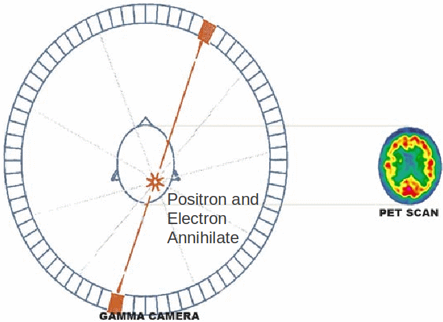
A positron is a subatomic particle - the antimatter particle to the electron. When positron and electron meet, they annihilate each other and the energy of both particles is released as two gamma rays travelling in opposite directions. The gamma rays must be emitted in opposite direction because only this way can momentum be conserved. The gamma rays can be used to indicate that electron and positron have annihilated, and the position of the annihilation can be deduced from the times at which the gamma rays are detected.
To take an image using a positron electron tomography scanner – also called a PET scanner, a patient drinks some water containing a positron emitting isotope with a short half life. A short half life isotope is needed so that the total does of radiation absorbed, and the danger to the patient is minimised. The liquid goes to each part of the body, emitting positrons, which annihilate with electrons to produce the pairs of gamma rays. The gamma rays are detected by a set of detectors which surround the patient on all sides, and used to build an image of the internal organs of the patient.
PET scanners are very expensive – much more expensive than ultrasound scans or X – rays. To be cost effective, a PET scanner must be in constant use and used only where other images will not produce an image that will help with a diagnosis.