With a simple x – ray photograph, it is hard to identify within soft tissue. There are two general techniques to take better images.
-
in a barium meal, a dense substance is swallowed, and the progress of the material along the gut is monitored.
-
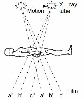
Tomography is a technique that makes the x – ray photograph focus on a certain region or slice through the patient, with all other regions blurred out of focus. This is achieved by moving the x – rays along with the film relative to the body as shown below.
An extension of basic tomography is the computerised tomography scan or CAT scan. This involves sending a pulse of x – rays and a set of sensitive detectors collects information about the level of radiation reaching each detector. The x – ray source and the detector are then rotated about the patient and the process repeated. A computer analyses the x – ray signals to construct a three dimensional image of the body.