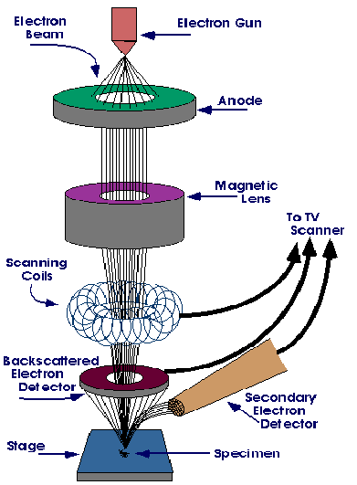
An electron microscope uses a particle beam of electrons to illuminate the specimen and produce a magnified image. Because resolving power is increases with decreasing wavelength, and electrons have wavelength 100000 times shorter than visible light, they can resolve down to 0.2nm and achieve magnifications up to 2000000x compared to 200nm and 2000x respectively for visible light.
The electron microscope uses electrostatic and electromagnetic "lenses" to control the electron beam and focus it to form an image. The electron beam is first diffracted by the specimen, and then, the electron microscope “lenses" re-focus the beam into an image.

Electron microscopes are used to image biological and inorganic specimens including microorganisms, cells, large molecules, biopsy samples, metals, and crystals and in industry for quality control in electronics.
The samples need not be crystalline and the processing of the image is easier than for X rays. But extremely thin sections of the specimens, typically less than 100 nanometers must be used for the electron microscope to give a good image. Biological specimens typically require to be chemically fixed, dehydrated and embedded in a polymer resin to stabilize them sufficiently to allow ultrathin sectioning. Sections of biological specimens, organic polymers and similar materials may require special `staining' with heavy atom labels in order to achieve the required image contrast.
