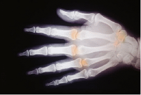
The basic principle of x – ray imaging is that some body tissues (especially bones) attenuate x – rays much more than other tissues. Photographic film darkens when a beam of x – rays are shone on them so bones show up as white areas on an x – ray picture.

Since x – rays cause ionisation, they are dangerous. This means they are taken only when necessary, and the x – ray dose is kept to a minimum. The dose may be reduced if the image can be enhanced by processing. There are two simple enhancement techniques:
-
When x – rays hit a screen the energy is partially re - radiated as visible light. The photographic film can absorb this extra light. The effect is to darken the image in the areas where the x – rays strike the film compared to the areas shield by the human body.
-
In an image intensifier tube, the x – rays strike a fluorescent screen and produce light, which then causes electrons to be emitted from a photocathode. The electrons are accelerated towards an anode where they strike another fluorescent screen and give off light to produce an image.
