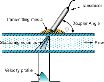
The doppler effect occurs when waves are emitted, reflected or detected by a moving object. The doppler effect can be used in medicine to analyse blood flow. When a pulse of ultrasound is sent along an artery, they are partially reflected by the blood cells. The blood cells are moving, so the reflected waves will display the doppler effect and the reflected waves will have different frequencies and wavelengths to the incident waves.
-
If blood is moving away from the ultrasound source, the wavelength of the reflected ultrasound will be increased and the frequency reduced.
-
If blood is moving towards the ultrasound source, the wavelength of the reflected ultrasound will be reduced and the frequency increased.
The greater the speed of the blood cells, the larger will be the difference between the frequencies (or wavelengths) of the incident and reflected waves. The speed of the the blood cells and be calculated from the magnitude of the doppler shift. Analysis of the doppler shift will display the pulsing movement of the blood as it is pumped by the heart, and can show whether the blood is flowing smoothly or if there is turbulence and the flow is being disrupted in some way. The presence of turbulence can mean that a blockage is developing, that the walls of blood vessels are weakening or that there is something wrong with the heart valves – maybe they are not opening or closing properly, impairing pumping efficiency or allowing leakage.
If the ultrasound is emitted at an angle![]() to the blood flow then the doppler shift is given by
to the blood flow then the doppler shift is given by![]()
where![]() is the speed of the blood and
is the speed of the blood and![]() is the speed of the ultrasound in blood.
is the speed of the ultrasound in blood.

Example: If![]() degrees, the doppler shift
degrees, the doppler shift![]() and the speed of ultrasound in blood is 1500 m/s then
and the speed of ultrasound in blood is 1500 m/s then![]()
