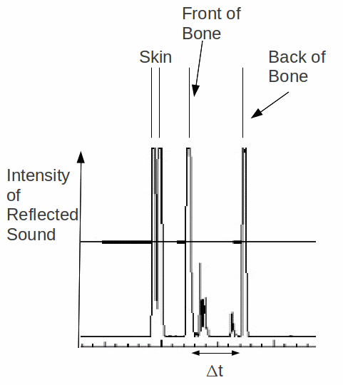
There are several types of ultrasound scan. One of the most common is the A – scan, often used for imaging the eye. It is a simple scan, in which a pulse of ultrasound is sent into the body and the reflected pulses are detected and displayed on an oscilloscope or computer screen as a voltage against time graph.

A pulse generator controls the ultrasound transducer. This pulse generator is connected to the time base of the oscilloscope. Simultaneously the pulse generator generates a pulse of ultrasound which travels into the patient. This starts a trace on the oscilloscope. Partial reflections are detected and displayed as a spike. The thickness of different tissues can be calculated and used to generate an image. The time interval between reflections from the front and back of bone above is![]() This is the time for wave reflected from the back of the bone to travel twice the thickness
This is the time for wave reflected from the back of the bone to travel twice the thickness![]() of the bone. If the speed of ultrasound in bone is
of the bone. If the speed of ultrasound in bone is![]() then
then![]()
Ultrasound is attenuated as it passes into the body, and the energy of the ultrasound is absorbed, so the reflections must be amplified.
