Ultrasound is a medical imaging technique that uses high frequency sound waves and their echoes. The technique is similar to echolocation used by bats, and dolphins, as well as SONAR and radar.
-
The ultrasound machine transmits high-frequency (1 to 5 megahertz) sound pulses into your body using a probe.
-
The sound waves travel into your body and hit a boundary between tissues (e.g. between fluid and soft tissue, soft tissue and bone).
-
Some of the sound waves get reflected back to the probe, while some travel on further until they reach another boundary and get reflected.
-
The reflected waves are picked up by the probe and relayed to the machine.
-
The machine calculates the distance from the probe to the tissue or organ (boundaries) using the speed of sound in tissue (5,005 ft/s or1,540 m/s) and the time of the each echo's return (usually on the order of millionths of a second).
-
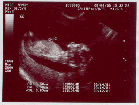
The machine displays the distances and intensities of the echoes on the screen, forming a two dimensional image like the one shown below of a foetus..

In a typical ultrasound, millions of pulses and echoes are sent and received each second. The probe can be moved along the surface of the body and angled to obtain various views.
The transducer probe is the main part of the ultrasound machine. It makes produces the sound waves and receives the echoes. The transducer probe generates and receives sound waves using a principle called the piezoelectric (pressure electricity) effect, which was discovered by Pierre and Jacques Curie in 1880. In the probe, there are one or more piezoelectric crystals. When an electric current is applied to these crystals, they vibrate producing sound waves Conversely, when sound or pressure waves hit the crystals, they emit electrical currents. Therefore, the same crystals can be used to send and receive sound waves. The probe also has a sound absorbing substance to eliminate back reflections from the probe itself, and an acoustic lens to help focus the emitted sound waves.
Transducer probes come in many shapes and sizes. The shape of the probe determines its field of view, and the frequency of emitted sound waves determines how deep the sound waves penetrate and the resolution.
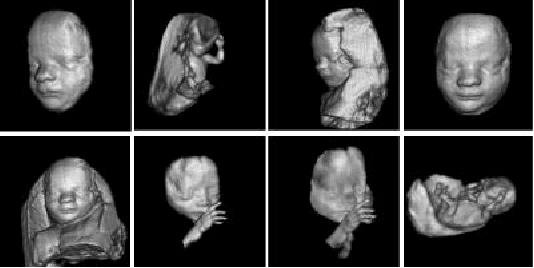
The ultrasound that we have described so far presents a two dimensional image, or "slice," of a three dimensional object (foetus, organ). Two other types of ultrasound are currently in use - 3D ultrasound imaging and Doppler ultrasound. Several two-dimensional images are acquired by moving the probes across the body surface or rotating inserted probes. The two-dimensional scans are then combined by specialized computer software to form 3D images, shown below.

3D imaging allows you to get a better look at the organ being examined and is best used for:
-
Early detection of cancerous and benign tumours
-
Visualizing a foetus to assess its development, especially for observing abnormal development of the face and limbs
-
Visualizing blood flow in various organs or a foetus
Doppler ultrasound is based upon the Doppler Effect. When the object reflecting the ultrasound waves is moving, it changes the frequency of the echoes, creating a higher frequency if it is moving toward the probe and a lower frequency if it is moving away from the probe. How much the frequency is changed depends upon how fast the object is moving. Doppler ultrasound measures the change in frequency of the echoes to calculate how fast an object is moving. Doppler ultrasound has been used mostly to measure the rate of blood flow through the heart and major arteries.
Ultrasound is safer and faster than X-rays or other radiographic techniques. Here is a list of uses:
-
measuring the size of the foetus to determine the due date
-
determining the position of the foetus to see if it is in the normal head down position or breech
-
checking the position of the placenta to see if it is improperly developing over the opening to the uterus (cervix)
-
seeing the number of foetuses in the uterus
-
checking the sex of the baby (if the genital area can be clearly seen)
-
checking the foetus's growth rate by making many measurements over time
-
detecting ectopic pregnancy, the life-threatening situation in which the baby is implanted in the mother's Fallopian tubes instead of in the uterus
-
determining whether there is an appropriate amount of amniotic fluid cushioning the baby
-
monitoring the baby during specialized procedures - ultrasound has been helpful in seeing and avoiding the baby during amniocentesis (sampling of the amniotic fluid with a needle for genetic testing). Years ago, doctors use to perform this procedure blindly; however, with accompanying use of ultrasound, the risks of this procedure have dropped dramatically.
-
seeing tumours of the ovary and breast
-
seeing the inside of the heart to identify abnormal structures or functions
-
measuring blood flow through the heart and major blood vessels
-
measuring blood flow through the kidney
-
seeing kidney stones
-
detecting prostate cancer early
In addition to these areas, there is a growing use for ultrasound as a rapid imaging tool for diagnosis in emergency rooms.
