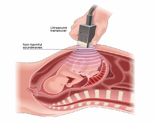
The upper limit of the frequencies that people can hear is 20,000 Hz. Any sound of higher freqency is known as ultrasound. Ultrasound used in medicine is typically just above 1 MHz. The velocity of sound in soft tissue is about 1500 m/s so the wavelength is of the order of a millimetre.
Unlike x – rays, ultrasound is not ionising so can safely be used to take images inside the body. A probe is used to emit and collect pulses of ultrasound reflected at boundaries between different types of tissue. There should be no air gap between probe and skin. If there were, almost all the ultrasound would be reflected. To ensure there is no air gap, gel or oil is spread over the skin, and the probe is moved over the gel or oil. The time taken between emitting and receiving pulses enables to work out distances and after processing, make images.

Very strong reflections can take place at the interface of tissues with very different densities, possibly causing multiple images by repeated reflecting between bones. The ultrasound probe is moved over the body to find a position at which these are minimised. The whole ultrasound scan can be captured on video for maximum viewing pleasure (my own witticism).
